
  |
|
表一 以MTT法測定黃麴毒素B1對鴨淋巴細胞毒害作用之試驗處理 Table 1. The experimental design of cytotoxic effect of aflatoxin B1 on duck lymphocytes by MTT assays |
|||||||||
| CTRL *- | CTRL+ | MIC | AFB1 104 ng/ml MIC | AFB1 104 ng/ml | AFB1 101 ng/ml MIC | AFB1 101 ng/ml | AFB1 10-2 ng/ml MIC | AFB1 10-2 ng/ml | |
| RPMI-1640
Lymphocytes (1×10⁷/ml) AFB1 PHA (200μg/ml) MIC (12.5 mg/ml) NADPH (0.25 mM) Total |
200
50
- -
-
-
250 |
175
50
- 25
-
-
250 |
170
50
- -
20
10
250 |
100
50
45 25
20
10
250 |
130
50
45 25
-
-
250 |
100
50
45 25
20
10
250 |
130
50
45 25
-
-
20 |
100
50
45 25
20
10
250 |
130
50
45 25
-
-
250 |
| *CTRL-=medium control;CTRL+=PHA alone;MIC=microsome alone;Volume unit=μl. | |||||||||
| 試驗四:培養系統加入不同最終濃度之β-胡蘿蔔素(0.025、0.05、0.1μM)和添加肝臟微粒體與黃麴毒素B1 (1×10⁴ ng/ml),最終細胞濃度為1×107淋巴細胞/ml,以探討黃麴毒素B1對鴨周邊血液淋巴細胞之致害作用及β-胡蘿蔔素對其是否具保護作用。試驗處理詳如表2。 |
|
表二 以 MTT 法 測 定 黃 麴 毒 素 B1 對 鴨 周 邊 血 液 淋 巴 細 胞 之 致 害 作 用 及 β- 胡 蘿 蔔 素 對 其 保 護 效 果 之 驗 處 理 Table 2. The experimental design of cytotoxic effect of aflatoxin B1 on duck lymphocytes and protective activity of β-carotene by MTT assays |
|||||||||
| CTRL- | CTRL+ | MIC | BCH | AFB1 104 ng/ml | AFB1 104 ng/ml MIC | AFB1 104 ng/ml MIC+ BCL | AFB1 104 ng/ml MIC+ BCM | AFB1 104 ng/ml MIC+ BCH | |
| RPMI-1640
Lymphocytes (1×10⁷/ml) AFB1 PHA (200μg/ml) betacarotene MIC (12.5 mg/ml) NADPH (0.25 mM) Total |
200
50
- -
- -
-
250 |
175
50
- 25
- -
-
250 |
170
50
- -
- 20
10
250 |
170
50
- -
30 -
-
250 |
130
50
45 25
- -
-
250 |
100
50
45 25
- 20
10
250 |
70
50
45 25
30 20
10
250 |
70
50
45 25
30 20
10
250 |
70
50
45 25
30 20
10
250 |
| *CTRL-=medium control;CTRL+=PHA alone;MIC=microsome alone;BCH=betacarotene alone (0.1μM);Volume unit=μl. | |||||||||
|
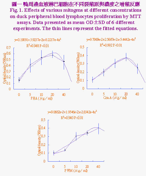
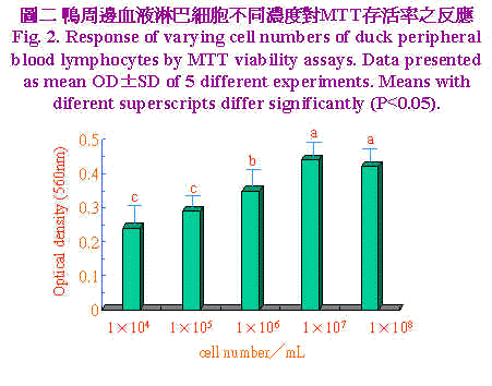
三、統計分析 實驗所得之資料先經一般線性模式(general linear model;GLM)進行變方分析,再以鄧肯氏新多項變域法(Duncan's new multiple range test)比較各組間差異顯著性。 結果與討論 不同裂殖原PHA、Con A及PWM於不同濃度(0、5、10、20和40μg/ml)加入培養系統中,以探討最佳MTT反應之裂殖原與濃度,結果示於圖1。資料經多項式(polynominal)迴歸所繪之迴歸曲線圖,由曲線圖可知裂殖原以PHA刺激增殖反應最佳,PWM次之,而ConA最差。迴歸方程式經一次微分求得增殖反應所須最大裂殖原濃度分別為PHA 23.86μg/ml、Con A 23.57 μg/ml和PWM 34.48 μg/ml。鴨周邊血液淋巴細胞不同濃度對MTT存活率之反應,示於圖2淋巴細胞數對MTT之反應呈二次曲線,當細胞濃度自1×104提高至1×107 cell/ml時,淋巴細胞數目持續增加其OD值亦升高,當細胞濃度再增加到1×108 cell/ml時OD值呈降低的趨勢,推測此一細胞濃度在經72小時培養後,營養分可能為其增殖之限制因子,亦即胎牛血清已用盡,由此一結果顯示以MTT方法測定鴨淋巴細胞之存活率以1×107 cell/ml最為適當,此一結果與Lessard and Dupuis (1994)試驗使用之細胞數一致。不同家畜禽之淋巴細胞表面之裂殖原受體數目與特性不一,對同一裂殖原反應時之最佳細胞數互異(Talebi et al., 1995)。 |


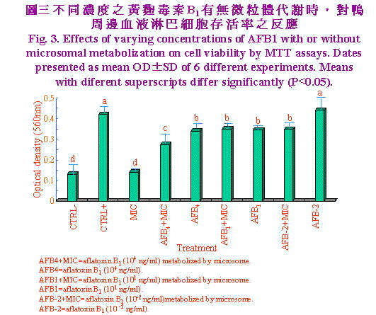
| 以不同濃度之黃麴毒素B1經有無微粒體代謝時,對鴨周邊血液淋巴細胞存活率之影響,結果示於圖3。單獨淋巴細胞或微粒體處理淋巴細胞增生數目相似,顯示微粒體對其並無刺激效果,但當PHA (20μg/ml)加入時,淋巴細胞受其刺激而大量增殖。黃麴毒素B1自高量(1×104 ng/ml)至低量時(1×101 ng/ml)OD值顯著(P<0.05)較單獨PHA處理者為低,此兩濃度顯然已造成淋巴細胞存活率之降低,但黃麴毒素B1 (1×10⁻² ng/ml)時對淋巴細胞增殖無抑制作用,可能為低量黃麴毒素且經淋巴細胞代謝後產生之毒性代謝物低至不影響其增殖作用。當高濃度黃麴毒素B1 (1×10⁴ ng/ml)經微粒體代謝後,顯著(P<0.05)抑制淋巴細胞增殖作用,由本試驗結果顯示黃麴毒素B1 (104 ng/ml)經代謝後可產生淋巴細胞致害作用,但濃度降低(1×101、1×10-2 ng/ml)時致害作用不顯著。 |

|
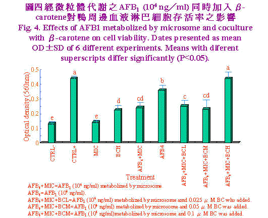
肝臟微粒體為細胞經均質化裂解而來,內質網斷裂後,再組合而成大小約100nm之小泡(Forrester et al., 1990),其富含多種酵素,其中以cytochrome p450在黃麴毒素B1代謝轉化為8,9-epoxide毒性增強(Ramsdell and Eation, 1990)。Neldon-Ortiz and Qureshi (1991)以經微粒體代謝之黃麴毒素B1作用雞腹腔巨噬細胞1小時,則發現微粒體使黃麴毒素B1毒性增強,甚至0.5μg/ml之濃度亦造成細胞形態改變,黏附力及吞噬能力亦明顯降低。相較本試驗使用之濃度104 ng/ml等於10 μg/ml,意即以淋巴細胞存活率來評估黃麴毒素B1經代謝之毒性上比以巨噬細胞之形態學、黏附力及吞噬力為評估指標較不敏感。Pang and Pan (1994)以豬淋巴細胞為對象,結果顯示黃麴毒素B1濃度於5×10⁴ ng/ml處理12小時,豬淋巴細胞死亡率即達50%。 經微粒體代謝之AFB1 (104 ng/ml)同時加入β-胡蘿蔔素(BC)對鴨周邊血液淋巴細胞存活率之影響,結果示於圖4。單獨微粒體處理與培養基對照組間無顯著差異,而當單獨高濃度BC (0.1 μM)添加時,OD值顯著(P<0.05)較培養基對照組為高,顯示BC為一潛在性裂殖原。單獨AFB1 (104 ng/ml)顯著(P<0.05)較AFB1經微粒體代謝者之細胞存活率為高,且中低濃度BC (0.05、0.025 μM)添加於AFB1經微粒體代謝之培養系統中時對細胞存活率並無改善之效果。然而高濃度BC (0.1 μM)添加於AFB1經微粒體代謝之培養系統中時極顯著的(P<0.05)較中低濃度BC者之細胞存活率為高,且改善至單獨PHA對照組相同之細胞存活率。 Chew et al., (1993b)實驗指出仔牛一次口服膠囊化BC 200 mg時,可提高淋巴細胞對BC之攝取量,但白血球及紅血球對BC攝取量無影響。進一步分析淋巴細胞之胞器。發現粒線體、胞核及微粒體中BC含量著增加,但仔牛血漿中BC及視覺醇含量無影響。本試驗之BC (0.1 μM)添加可有效的提高淋巴細胞存活率,可能與提高淋巴細胞對BC之攝取有關;雖然對於BC在調節免疫力上所扮演的角色尚不瞭解,但BC可能以自由基清除者抵抗自由基導致之細胞受損。事實上,生體內和生體外研究指出,BC可促進淋巴細胞增殖作用(Hoskinson et al., 1989)及增加助手T細胞數目(Prabhala et al., 1989)。而本試驗之淋巴細胞含高濃度BC可能保護或對抗AFB1經代謝後產生之自由基,此外BC也可能直接調節淋巴細胞之增殖。Chew et al., (1993b)實驗發現視覺醇並未隨之升高,顯示BC有別於一般人認為僅是維生素A原之刻板印象,而是有其獨特的作用。 雖然AFB1造成淋巴細胞死亡的確實機制未知,但AFB1經微粒體酵素代謝之二次產物造成細胞膜不穩定可能扮演一重要地位(Pokrovsky et al., 1972)。曾有報告指出,氧自由基造成細胞膜受損,而致脂質過氧化、蛋白質及DNA之破壞(Perera et al., 1987)。近來研究顯示AFB1及其代謝物可經酵素非酵素作用形成自由基(Kodama et al., 1990),是否自由基為造成淋巴細胞存活率降低之主因,須加以進一步證實。Lessard and Dupuis (1994)生體試驗也陳述日糧中BC含量增加,雞淋巴細胞增殖作用及自然殺手細胞(natural killer)活性亦提高。但亦有試驗指出BC或BC與維生素E協同時無法提高以E. coli免疫雞隻的凝集抗體力價,維生素E或A卻可以提高,此可能與BC對疾病保護機制異於維生素E或A有關。 生體外試驗,當添加BC濃度為10-8M對鼠脾臟淋巴細胞增殖無影響,添加BC10-5M抑制牛週邊血液淋巴細胞之增殖作用(Tjoelker et al., 1988),此一結果與本試驗結果相去甚遠,其原因可能為不宣品種動物對BC攝取程度差異及free radical存在與否時造成之影響。此一推論基礎可由Lawlor and O'brien (1995)應用paraquat 0.25 mM處理雞胚纖維母細胞(chicken embryo fibroblast, CEF)18小時,造成其氧化性緊迫,當0.1μM BC添加時,superoxide dimutase及glutathione peroxidase (GSH-px)活性回復至與對照組相同水準,且降低catalase活性。當10μM BC添加時superoxide dimutase及catalase顯著升高且GSH-px活性降低至與對照組相當。顯示BC有效保護CEF對抗paraquat誘發之氧化性緊迫。 綜觀本試驗結果顯示,高濃度AFB1 (104 ng/ml)經代謝後對鴨淋巴細胞毒性增強,高濃度胡蘿蔔素(0.1 μM)可保護鴨淋巴細胞免除此一毒害作用,是否為抑制氧自由基之產生,須進一步探討。 |

|
參考文獻 鄭永祥、龐飛。1995。黃麴毒素B1對鴨淋巴細胞轉型作用之影響。中華農學會報170:147~155。 Bendich, A., and S. S. Shapiro. 1986. Effect of β-carotene and canthaxanthin on the immune responses of the rat. J. Nutr. 116: 2254~2261. Bendich, A. 1991. Carotenoids and immunity Clin. App. Nutr. 1: 45~53. Bounous, D. I., R. P., Campagnoli and J. Brown. 1992. Comparoson of MTT colorimetric assay and titrated thymidine uptake for lymphocyte proliferation assay using chicken splenocytes. Avian Disease 36: 1022~1027. Chew, B. P., 1993a. Role of carotenoids in the immune response. J. Dairy Sci. 76: 2804~2811. Chew, B. P., T. S. Wong and J. J. Michal. 1993b. Uptake of orally administered β-carotene by blood plasma, leukocytes, and lipoproteins in calves. J. Anim. Sci. 71: 730~739. Denizot, F. and R. Lang. 1986. Rapid colorimetric assay for cell growth and survival modification to the tetrazolium dye procedure giving improved sensitivity and reliability. J. Immunol. Method 89: 271~277. Forrester, L. M., G. E. Neal, D. J. Judah, M. J. Galancey and C. R., Wolf. 1990. Evidence of involvement of multiple forms of cytochrome P-450 in aflatoxin B1 metabolism in human liver. Proc. Natl. Acad. Sci. 87:8306~8310. Hoskinson, C. D., B. P. Chew and T. S. Wong. 1989. Effects of β-carotene and vitamin A on mitogen-induced lymphocyte proliferation in the pig in vivo. FASEB J3:A663. Kadian, S. K., D. P. Monga and M. C. Goel. 1988. Effect of aflatoxin B1 on the delayed type hypersensitivity and phagocytic activity of reticuloendothlial system in chickens . Mycopathologic 104 :33~36. Kodama, M., F. Inoue and M. Akao. 1990. Enzymatic and non-enzymatic formation of free radicals from aflatoxins B1. Free Rad. Res. Comms. 10: 137~142. Lawlor, S. M. and N. M. O'brien. 1995. Modulation of oxidative stress by β-carotene in chicken embryo fibroblasts. Brit. J. Nutr. 73: 841~850. Lessard, M. and M. Dupuis. 1994. Differential modulation of chicken lymphocyte blastogenesis and cytotoxic activity of natural killer cells in vitro by retinol, retionic acid and beta-carotene. Nutr. Res. 14:1201~1217. Lowry, O. H., N. J. Rosebrough, A. L. Far and R. J. Randall. 1951. Protein measurement with folin phenol reagent J. Biol. Chem. 193: 265~275. Mosmann, T. 1983. Rapid colorimetric assay for cell growth and survival: Application to proliferation and cytotoxicity assays. J. Immunol. Method 65:55~63. Neldon-Ortiz, D. L. and M. A. Qureshi. 1991. Direct and microsomeal activated aflatoxin B1 exposure and its effects on turkey peritoneal macrophage functions in vitro. Toxicol. Appl. Pharmacol. 109: 432~442. Pang, V. F. and C. Y. Pan. 1994. The cytotoxic effects of aflatoxin B1 on swine lymphocytes in vitro. J. Chin. Soc. Vet. Sci. 20(4):289~301. Perera, M. I. R., J. M., Bestschart, M. A. Virji, S. L. Katyal, H. Shinozuka. 1987. Free radical injury and liver tumor promotion. Toxicol. Pathol. 15:51~59. Peterson, G. L. 1977. A simplification of the protein assay method of Lowery et al. which is more generally applicable. Anal. Biochem. 83: 346~356. Pier, A. C., R. E. Fichtner and S. J. Cysewski. 1977. Effect of aflatoxin on the cellular immune system. Ann. Naer. Alim. 31:781~788. Pokrovsky, A. A., L. V. Kravchenko and V. A. Tutelyan. 1972. Effect of aflatoxin on rat liver lysosomes. Toxicon. 10:25~30. Prabhala, R. H., V. Maxey, M. J. Hicks and R. R. Watson. 1989. Enhancement of the expression of activation markers of human peripheral blood mononuclear cells by in vitro culture with retinoids and carotenoids. J. Leukocyte Biol. 45: 249~256. Pamsdell, H. S. and D. L. Eaton. 1990. Species susceptibility to aflatoxin B1 carcinogenesis: Comparative kinetics of microsomal biotrans formation. Cancer Res. 50: 615~620. Talebi, A., P. R. Torgerson and G. Mulcahy. 1995. Optimal conditions for measurement of blastogenic reponses of chickens to concanavalin A in whole blood assays. Vet. Immunol. and Immunopathol. 46: 293~301. Tengerdy, R. P., N. G. Lacetera and C. F. Nockels. 1990. Effect of beta carotene on disease protection and humoral immunity in chickens. Avian disease 34: 848~854. Tjoelker, L. W., B. P. Chew, T. S. Tanaka and L. R. Daniel. 1988. Bovine vitamin A and β-carotene in take and lactational status. 2. Responsiveness of mitogenstimulated peripheral blood lymphocytes to vitamin A and β-carotene challenge in vitro. J. Dairy Sci. 71: 3120~3127. |
|
The Cytotoxic Effect of Aflatoxin B1 on Duck Lymphocytes and the Protective Effect of β-Carotene Yeong-Hsiang Cheng(1) and Victor-Fei Pang(2) Received Oct. 26, 1995; Accepted Jan. 29, 1996 ABSTRACT The purpose of this experiment was to determine duck peripheral blood lymphocyte proliferation by a MTT (3-(4,5-dimethyl thiazol-2-yl)-2,5-diphenyltetrazolium bromide) assay. This assay was also used to evaluate the cytotoxic effect of aflatoxin B1 (AFB1) on duck lymphocyte and the protective effect of β-carotene (BC). Trial 1: Three mitogens were examined and the results showed that phytohaemagglutinin (PHA) had the best proliferation response, pokeweed mitogen (PWM) intermediate, and Concanavalin A (Con A) the least. Using best-fit regression, the best mitogen concentrations for proliferation response were PHA, 23.68 μg/ml;Con A, 34.48μg/ml and PWM, 23.57 μg/ml, respectively. Trial 2: The proliferation response of lymphocyte number by a MTT assay showed a quadratic fashion. When cell numbers were raised from 1×10⁴ to 1×10⁷ cell/ml, the OD values were also increased. However, the OD values tended to decrease when cell numbers were increased to 1×10⁸ cell/ml. Trial 3:The results showed that AFB1 treatment from high dose (1×10⁴ ng/ml) to low dose (1×101 ng/ml) had OD values significantly lower (P<0.05) than PHA treatment alone. But, there was no inhibitory effect when AFB1 Aat an extremly low dose (1×10⁻² ng/ml) was introduced. High dose of AFB1 which was metabolized by microsome inhibited lymphocyte proliferation to a greater extent than AFB1 which was without microsome metabolization. There was no significant inhibition at low dose (1×101,1×10-2 ng/ml) treatments. Trial 4: The results showed that when high BC level (0.1μM) was added, OD values of lymphocyte proliferation were higher (P<0.05) than medium control. Treatment of AFB1 alone had higher (P<0.05) proliferation than AFB1 metabolized by microsome. There was no improvement on lymphocyte proliferation when medium and low levels of BC (0.05, 0.025 μM) were added to culture system of AFB1 metabolized by microsome. However, when high level of BC was added to the same culture system, the proliferation was higher (P<0.05) than medium and low levels of BC and the improvement was the same as PHA control group. This experiment revealed that high dose of AFB1 metabolized by microsome had an intensified effect on inhibition of duck lymphocyte proliferation. However, high levels of BC could alleviate this detrimental effect on duck lymphocyte. (Key Words: Duck, Aflatoxin B1, Lymphocyte, β-Carotene) ─────────────────────────────────── (1)Department of Animal Science, National 1-Lan Institute of Agriculture and Technology, I-Lan, Taiwan, R.O.C. (2)Department of Veterinary Medicine, National Taiwan University, Taipei, Taiwan, R.O.C. |